
Seegene

Technology

Seegene High
Multiplex
PCR Technology

High Multiplex Detection in One Test​

STST™

Single Temperature
Single Target

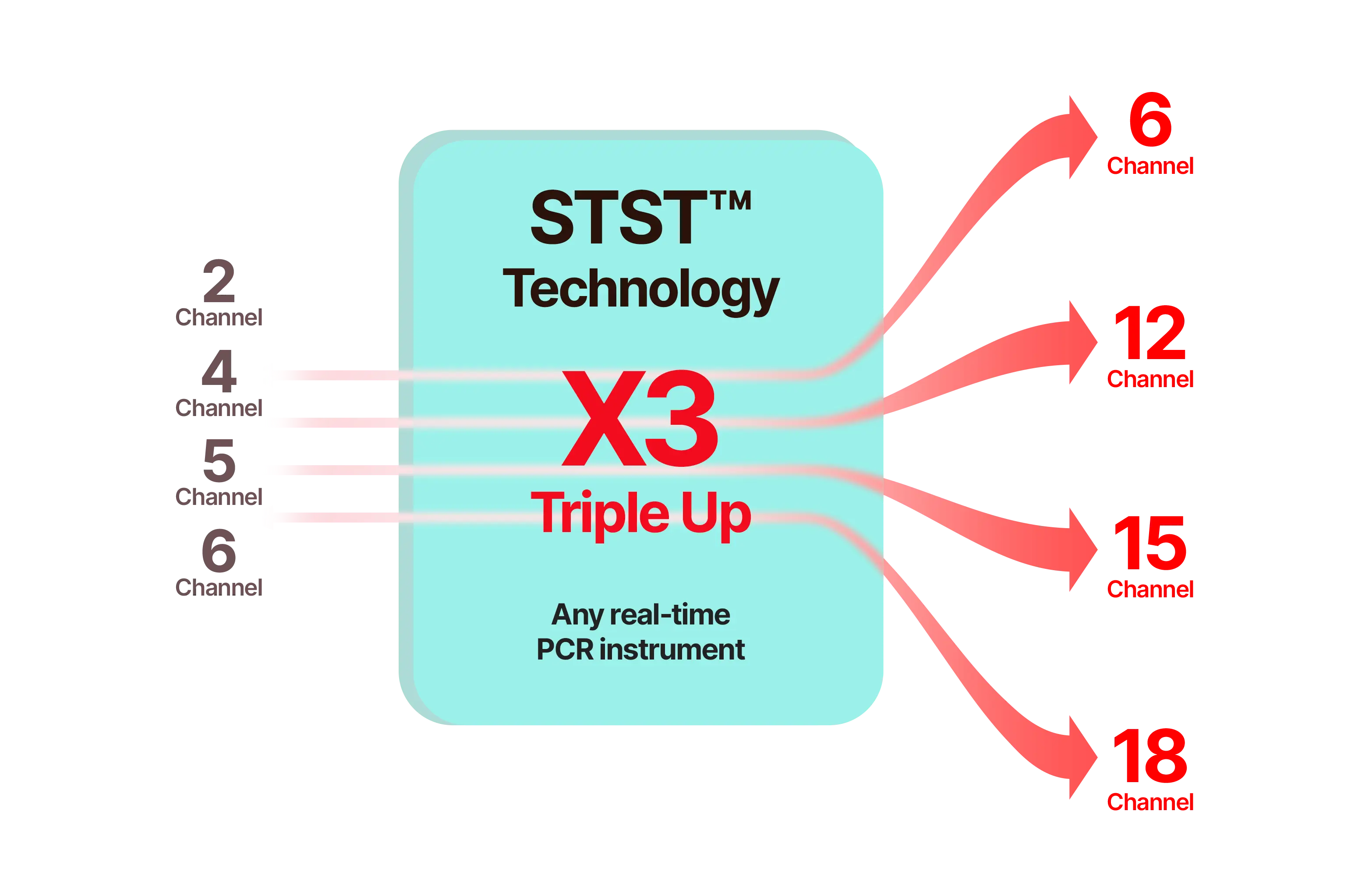
STST™ is a next-generation PCR reagent platform designed to generate independent, non-overlapping fluorescent signals at a single temperature. This structural signal separation eliminates the need for post-processing required in conventional multiplex PCR, enabling accurate quantification of up to five targets within a single channel. With a simplified analytical workflow and intuitive signal interpretation, STST™ improves both the speed and precision of diagnostics while maximizing multiplexing performance across various instrument environments.​

STST™ Features
  1. Quantitative multiplexing within a single channel​

    • Detects up to five targets in one channel​
    • Provides independent, non-overlapping signals without post-run processing​
  2. Direct and reliable signal interpretation​

    • “What you see is what it is” — stable signals directly reflecting each target​
    • No additional analysis or deconvolution required for Ct calculation
  3. Expanded effective multiplexing capability​

    • Overcomes hardware limits to deliver enhanced multiplexing performance​
    • Enables multi-target quantification even on single-channel instruments​
  4. Faster decision-making with a simplified workflow​

    • Clear signal profiles allow rapid interpretation and reporting​
    • Reduced analysis time supports quicker clinical decisions
Principle of STST™ technology​

STST generates a single signal for each temperature, achieving 5Ct detection and 25plex performance.​

A comparison chart of MuDT™ and STST™ technologies. MuDT™ shows how targets A, B, and C are detected across different temperature stages (83°C, 72°C, 60°C), with cumulative RFU signals displayed for each plotting step. STST™ demonstrates direct target detection at each temperature, showing separate amplification curves for A, B, and C without signal overlap.
STST™ TECHNOLOGY X3 TRIPLE UP​​

A simple way to triple multiplexing performance ​on any real-time PCR instrument

STST™ generates clean, independent fluorescence signals at a single temperature, allowing any existing real-time PCR instrument to triple its multiplexing capability without hardware upgrades.​ With interference-free signals and the structural advantages of STST™, signal noise is reduced and reproducibility is improved, providing reliable and faster diagnostic results without complex post-processing.

Diagram showing STST™ Technology X3 Triple Up expanding 2–6 PCR channels into 6–18 channels using clean, independent fluorescence signals at a single temperature.

The Next Generation Real-time PCR

MuDT™

Multiple Detection Temperatures

MuDT™ technology is the analytical real-time PCR technology detecting two targets with individual Ct values in a single channel without melting curve analysis. By utilizing differentiated fluorescence signals generated at two distinct detection temperatures, MuDT™ enables accurate Ct value determination for each pathogen, even in co-infected samples. Combined with DPO™ and TOCE™, MuDT™ significantly enhances the ability to deliver high-multiplex, clinically actionable diagnostics that improve patient care while reducing healthcare costs.

MuDT™ Features
  1. Multiplexing with Ct values in a single channel

    • Detecting two targets in a single channel
    • Providing Ct values for each target
  2. The detection and quantification for two targets in a single reaction

    • Comprehensive information for improved patient care
  3. Each Ct value of co-infected pathogens equals that of single target amplification

Building on this foundation, Seegene developed 3 Ct™ technology, which expands multiplexing capacity from two to three independent targets per fluorescent channel, enabling simultaneous detection and relative quantification. This advancement dramatically elevates multiplexing performance while maintaining speed, accuracy, and cost-effectiveness.

3 Ct™ Technology

3 Ct™ technology is a breakthrough that combines Seegene’s proprietary technologies specialized in high multiplexing with advanced analysis algorithms. It allows the precise differentiation of three different targets in a single fluorescent channel, providing individual Ct values for each target and enabling relative quantification. It also achieves rapid amplification by applying fast enzyme and significantly reduces turnaround time by analyzing Ct values instead of melting curves.

3 Ct™ technology advances the field of multiplex PCR by enabling real-time detection of up to 15 targets with individual Ct values in a single reaction, delivering true high-multiplex PCR performance that supports accurate and competitive syndromic testing

3 Ct™ Features
  1. Maximized multiplexing with Ct value

    • Detection of up to three targets in a single channel
    • Relative quantitative information for each target
  2. Short turnaround time

    • Achieved faster results in just 1.5 hours
    • No additional analysis required
  3. Cutting-edge analysis algorithm for reliable and accurate results

    • Implemented with Seegene’s 19 core patents
    • Integrated with Seegene Viewer for automated analysis
  4. Competitive syndromic testing

    • Individual detection of 15 targets from a single reaction
    • High throughput and cost-effectiveness
    • Expandable for diagnosis of various diseases
Principle of 3 Ct™ technology

Fluorescence signals are measured at different temperatures to provide multiple Ct values in a single channel. The amount of signal at each point is differentially analyzed and displayed in Seegene Viewer.

Tm=83°C, Tm=83°C, Tm=83°C. Seegene Viewer. Directly use 83°C, 1st plotting 72°C–83°C, 2nd plotting 60°C–72°C–83°C. Result for A (High Tm target), Result for B (Mid Tm target), Result for C (Low Tm target).
Seegene assay has unique advantages based on its advanced multiplex technology
Seegene vs Other Companies. Seegene’s Syndromic Technology: 14 Targets + IC, 15-plex in one tube, 3 Ct™ in a channel, 19 patents applied, Syndromic assay* (*Comprehensive and simultaneous testing for all potential causative pathogens). Other Companies’ Conventional Technology: 3 Targets + IC, 3 Targets + IC, 3 Targets + IC, 3 Targets + IC, 2 Targets + IC, 4-plex in one tube, 1 Ct in a channel, TaqMan applied, Targeted assay* (*Selective testing for suspicious causative pathogen).
Example of analysis results - Allplex™ HPV HR Detection
Three RFU vs Cycle graphs for HPV66, HPV45, and HPV58. Graph 1: HPV66, Ct 19.31. Graph 2: HPV45, Ct 36.93. Graph 3: HPV58, Ct 36.76. Below graphs, a result table shows: Type - SAMPLE; Auto Interpretation - HPV66, 45, 58; FAM Channel columns - HPV66 (Ct 19.31, positive), HPV45 (Ct 36.93, positive), HPV58 (Ct 36.76, positive).

New Paradigm of Multiplex Real-time PCR

TOCE™

Tagging Oligonucleotide Cleavage and Extension

TOCE™ technology is a very elegant oligonucleotide chemistry solution for the homogenous high multiplex real-time PCR. TOCE™ technology overcomes the technical limitations of target-based probe real-time PCR technology and fully exploits the potential of real-time PCR in high multiplex analysis, through unique signal generation & melting temperature analysis by novel components.

TOCE™ technology also supports the detection and distinction of multiple targets in a single tube and provides quantitative results through cyclic-CMTA analysis. This capability forms the foundation of Seegene’s single-channel high-multiplex PCR platform, allowing scalable and efficient multi-target detection.

TOCE™ Features
  1. High multiplicity in a single channel using Catcher-Tm analysis

  2. Controllable Catcher-Tm profile

  3. Consistent Catcher-Tm values regardless of target sequence variations

  4. Sensitivity comparable to singleplex real-time PCR

  5. Multiple quantitative analyses using cyclic-CMTA in a single channel

  6. High multiple point-mutation detections in a single tube

Multiple detection in a single channel

Multiple detection in a single channel. A red line graph showing multiple distinct peak signals within one fluorescence detection channel, indicating the ability to detect multiple targets from a single reaction curve.
Principle of TOCE™ technology

The key components for TOCE™ technology are DPO™ primer pairs, Pitchers and Catchers. The DPO™ provides highly specific amplification of the target region. The Pitcher is a tagging oligonucleotide which hybridizes specifically to the target region. The Catcher is a dual-labeled artificial template.The key components for TOCE™ technology are DPO™ primer pairs, Pitchers and Catchers. The DPO™ provides highly specific amplification of the target region. The Pitcher is a tagging oligonucleotide which hybridizes specifically to the target region. The Catcher is a dual-labeled artificial template.

The 5’ nuclease activity specifically cleaves a target-bound Pitcher and then the designed tagging portion is released. The released tagging portion hybridizes with the capturing portion of the Catcher. The duplex catcher formation induces extension on the Catcher, thus resulting in the generation of a fluorescence signal. The signal can be analyzed in manners of real-time or melting curve analysis.

DPO™ mechanism diagram showing the Pitcher primer binding first and guiding the Catcher primer. Three catcher probes are shown with different melting temperatures: Tm=70°C with green catcher probe and quencher, Tm=65°C with blue catcher probe and quencher, and Tm=60°C with red catcher probe and quencher. Each catcher includes a reporter (R) and quencher (Q) for target-specific fluorescence detection.
Three peak curves representing catcher melting temperatures. Red peak at 60°C, blue peak at 65°C, and green peak at 70°C, labeled as Catcher Tm.
Principle of quantitative analysis by cyclic-CMTA

In the left panel, CMTA points, preselected cycles during the amplification process where melting temperature analysis is performed, are indicated (1, 2, 3). The right panel shows the appearance of the melting peak for three different titers of the target. The melting peak appears at the first CMTA point for the high titer, at the second CMTA point for the intermediate titer, and at the third CMTA point for the low titer.

Amplification curve graph. X-axis shows cycles from 0 to 50 and Y-axis shows RFU from 0 to 3000. A turquoise amplification curve rises from baseline around cycle 30, increases steeply around cycle 40, and plateaus near cycle 50. Three labeled points are marked: 1 at the beginning of the rise, 2 during the steep increase, and 3 at the plateau.
Table showing cyclic-CMTA points across 1st (30 cycles), 2nd (40 cycles), and 3rd (50 cycles) at 65°C, with interpretation by titer level. High titer shows peak curves at all three cycle points and is interpreted as +++. Intermediate titer shows a peak only at the 2nd and 3rd cycles and is interpreted as ++. Low titer shows a peak only at the 3rd cycle and is interpreted as +. Not detected shows no peaks at any cycle and is interpreted as Not detected.

Superiority of Multiplex PCR

DPO™

Dual Priming Oligonucleotide

DPO™ technology is the innovative multiple target amplification technology that enhances target specificity and minimizes the non-specific amplification that commonly occurs in multiplex PCR. DPO™ technology redefines high multiplex PCR by enabling the detection of “many targets in a single tube” and is well-suited for high multiplex real-time PCR technology.

Principle of DPO™ technology

DPO™ primer is structurally different from the conventional primer and is composed of two functional priming portions connected by the polydeoxyinosine (Poly-d(I)) linker. The length of the 5’-end target stabilizing portion is longer than that of the 3’-end target determinant portion. Poly-d(I) linker forms a bubble-like structure at a certain annealing temperature and controls the two-step priming reactions required for the polymerase to extend a DPO™ primer. With a DPO™ primer, initial binding is through the 5’-end portion, and then the primer extension is controlled through more specific annealing of the 3’-end portion.

Diagram of DPO primer structure showing a bubble-like Poly-d(I) linker between a 5′ stabilizer region and a 3′ determiner region. Step 1: The long 5′-end portion binds the target DNA to initiate stable annealing. Step 2: The short 3′-end portion selectively binds to the target site and determines target-specific extension.
Unparalleled specificity by eliminating any false extension

DPO™ primer does not generate any false positive signal by blocking the extension of primer on a non-target template, thus provides unparalleled specificity in high multiplex PCR.

Two diagrams comparing non-specific binding prevention. A: The longer 5′-end portion binds a non-target site, but the short 3′-end portion prevents non-specific extension. B: The short 3′-end portion alone fails to bind at the annealing temperature, preventing extension at non-target sites.
Example (Comparison of Multiplex PCR results with DPO™ primer vs. conventional primer)

DPO™ primer evaded non-specific amplification and produced only true target bands.
However, conventional primer did not avoid nonspecific amplification.

DPO™ multiplex PCR assay is the most accurate, rapid, and cost-effective method.

Gel electrophoresis comparison of Conventional primers vs DPO primers. The conventional primer gel (lanes N, 1–5, P) shows multiple non-specific bands. The DPO primer gel (lanes N, 1–5, P) shows clearer and more specific bands corresponding to Flu A, Flu B, RSV B, RSV A, and OC43 targets.Gel electrophoresis comparison of Conventional primers vs DPO primers. The conventional primer gel (lanes N, 1–5, P) shows multiple non-specific bands. The DPO primer gel (lanes N, 1–5, P) shows clearer and more specific bands corresponding to Flu A, Flu B, RSV B, RSV A, and OC43 targets.
Explore Seegene's comprehensive portfolio of diagnostic solutions
View more